Category:Biology images that should use vector graphics

Images are placed here by a adding {{Convert to SVG|biology}} to the image page.
Subcategories
This category has the following 3 subcategories, out of 3 total.
Pages in category "Biology images that should use vector graphics"
This category contains only the following page.
Media in category "Biology images that should use vector graphics"
The following 200 files are in this category, out of 1,690 total.
(previous page) (next page)-
-
(zh)Ips cells.png 629 × 600; 145 KB
-
0321 DNA Macrostructure.jpg 743 × 584; 227 KB
-
1008 Skeletal Muscle Contraction.jpg 811 × 1,259; 478 KB
-
1015 Types of Contraction new.jpg 741 × 1,083; 286 KB
-
1028 Smooth Muscle Contraction.jpg 1,126 × 233; 139 KB
-
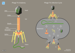
11 Hegasy Phage T4 Wiki E CCBYSA.png 2,109 × 1,476; 193 KB
-
12 Hegasy Cas9 Immun Wiki D CCBYSA.png 2,082 × 1,476; 268 KB
-
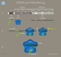
13 Hegasy CRISPR pre crRNA Wiki D CCBYSA.png 2,074 × 1,943; 268 KB
-
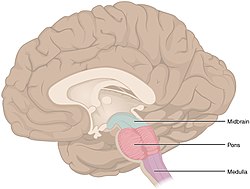
1311 Brain Stem.jpg 906 × 685; 208 KB
-
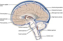
1317 CFS Circulation.jpg 1,065 × 693; 373 KB
-
 15010 2020 1486 Fig3 HTML.webp 1,502 × 612; 91 KB
-
1503 Connections of the Parasympathetic Nervous System.jpg 1,967 × 3,021; 676 KB
-
1506 Referred Pain Chart.jpg 1,983 × 1,392; 704 KB
-
16S and 28S phylogenies of three Waldo species - ZooKeys-316-067-g003.jpeg 1,512 × 750; 222 KB
-
1756-8935-5-4-3-l.jpg 1,200 × 1,488; 227 KB
-
1806 The Hypothalamus-Pituitary Complex.jpg 1,077 × 626; 248 KB
-
1axc PCNA ProCheck Rama.jpg 500 × 500; 158 KB
-
1ملف.png 330 × 379; 90 KB
-
2008 Internal Anatomy of the HeartN.jpg 1,656 × 1,159; 760 KB
-
201012 Vero.png 408 × 433; 188 KB
-
201405 cerebellem.png 400 × 400; 62 KB
-
201405 mammary gland.png 400 × 400; 70 KB
-
201710 Cells.png 1,000 × 1,000; 1.42 MB
-
2032 Automatic Innervation.fr.jpg 1,110 × 2,071; 413 KB
-
3D medical animation coronavirus structure.jpg 2,150 × 1,000; 1.14 MB
-
3rd gen Epigenetics.png 1,077 × 493; 77 KB
-
502 Layers of epidermis (no labels).png 792 × 647; 563 KB
-
502 Layers of epidermis.jpg 1,080 × 854; 656 KB
-
5fold 2dom 3CDX ribbon.png 900 × 900; 372 KB
-
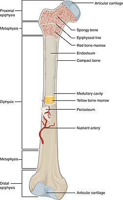
603 Anatomy of Long Bone.jpg 708 × 1,156; 257 KB
-
604 Bone cells.jpg 807 × 567; 198 KB
-
709 2017 1147 Fig3c.jpg 620 × 618; 60 KB
-
800px-HIV Viron es.png 2,800 × 2,135; 1.78 MB
-
A range of putative disease-causing mechanisms for the case of HGPS.jpg 1,200 × 874; 221 KB
-
A textbook of obstetrics (1899) (14591591600).jpg 1,848 × 1,308; 129 KB
-
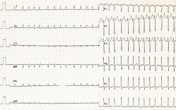
A-Fib-large.jpg 710 × 572; 289 KB
-
A-Fib.jpg 406 × 327; 113 KB
-
A-fib.png 1,539 × 757; 2.24 MB
-
Abb. 4 Ökologische Mykorrhiza-Module.tif 3,346 × 2,574; 1.09 MB
-
Abdominal hair patterns cmg.jpg 461 × 640; 58 KB
-
ACh Rezeptor Konformation.png 734 × 396; 25 KB
-
ACh rezeptor.png 734 × 664; 35 KB
-
ACh-Rezeptor Mechanism.png 814 × 606; 48 KB
-
Acid-sec-ru.PNG 987 × 1,194; 3.38 MB
-
Acidose metabólica com hiato aniônico elevado.png 1,103 × 731; 36 KB
-
Actin myosin filaments.png 720 × 315; 50 KB
-
Actinomyxon zh hans.png 644 × 1,092; 9 KB
-
Action potential ventr myocyte.gif 960 × 720; 25 KB
-
Active zone3.JPG 876 × 796; 108 KB
-
Adenovirus structure.png 800 × 400; 95 KB
-
ADN-ARN.png 189 × 275; 39 KB
-
Adrenozeptor-Signaltransduktion.jpg 538 × 580; 54 KB
-
Adressage à la membrane du récepteur GABAB.png 2,503 × 1,875; 669 KB
-
Adult and baby skin.jpg 1,240 × 1,470; 169 KB
-
Adultvocaltract.jpeg 377 × 500; 127 KB
-
Aegicetus CGM 60584 skeleton.png 6,272 × 1,444; 1.2 MB
-
Aeolidiella stephanieae 12.png 672 × 645; 146 KB
-
Aeolidiella stephanieae 13.png 651 × 635; 130 KB
-
Aeolidiella stephanieae 14.png 677 × 658; 139 KB
-
Aerobic mitochondria process.png 576 × 376; 48 KB
-
Aerobic pathways.png 432 × 597; 39 KB
-
Aerobic production pathways.png 864 × 255; 36 KB
-
Afferents.png 1,331 × 1,326; 139 KB
-
Afib ecg (CardioNetworks ECGpedia).jpg 2,000 × 987; 475 KB
-
Afps 3ptcpr.png 633 × 229; 18 KB
-
Afps ptcpr carotid.png 352 × 217; 8 KB
-
Afps ptcpr femor.png 455 × 290; 29 KB
-
Afps ptcpr humer.png 327 × 193; 9 KB
-
Alanine Scan.png 1,075 × 958; 351 KB
-
AlgaeTree.png 540 × 619; 8 KB
-
Alley 01.jpg 725 × 279; 5 KB
-
Alley 02.jpg 727 × 277; 11 KB
-
Alley 03.jpg 727 × 274; 10 KB
-
Alley 04.jpg 727 × 277; 15 KB
-
Alley 05.jpg 720 × 275; 11 KB
-
Alley 06.jpg 726 × 277; 19 KB
-
Alley 07.jpg 712 × 272; 12 KB
-
Alley 08.jpg 725 × 274; 8 KB
-
Alley 10.jpg 734 × 274; 13 KB
-
Alley 11.jpg 725 × 273; 10 KB
-
Alley 12.jpg 730 × 277; 17 KB
-
Alley 13.jpg 730 × 277; 13 KB
-
Alley 14.jpg 727 × 274; 10 KB
-
Alley 15.jpg 727 × 274; 10 KB
-
Alley 16.jpg 727 × 274; 15 KB
-
Alley 17.jpg 727 × 274; 9 KB
-
Almohadillas.jpg 473 × 205; 19 KB
-
AlphaHemolysin membrane heptamer 7AHL VwDownPore.jpg 1,600 × 1,602; 1.06 MB
-
Alternative splicing.jpg 2,180 × 1,726; 625 KB
-
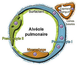
Alveole.jpg 363 × 304; 46 KB
-
Alveoli diagram.png 569 × 477; 23 KB
-
Alzheimer.jpg 482 × 322; 63 KB
-
Amazona vittata range.PNG 2,058 × 642; 56 KB
-
Ambulocetus.png 800 × 263; 37 KB
-
Amigdale1.jpg 197 × 185; 29 KB
-
Amino acid catabolism (previous version).png 1,838 × 1,204; 250 KB
-
Amniotic fluid embolism.png 2,488 × 3,504; 1.61 MB
-
Amy1 gene in apes.png 919 × 551; 53 KB
-
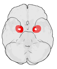
Amyg.png 189 × 230; 22 KB
-
Amygdala dorsal view.png 767 × 932; 430 KB
-
Amygdala frontal view.png 1,026 × 1,007; 597 KB
-
Amygdala left lateral slice.png 939 × 763; 385 KB
-
Amylopectine2.PNG 845 × 500; 49 KB
-
Amylose2.PNG 590 × 418; 32 KB
-
 An overview of COVID-19 therapeutics and drugs.webp 1,960 × 1,188; 194 KB
-
 An overview of designing and developing COVID-19 vaccines.webp 1,770 × 1,770; 294 KB
-
ANA - dsDNA antibody.png 492 × 494; 18 KB
-
ANA Immunofluorescence.png 720 × 340; 28 KB
-
Anagen catagen telogen.jpg 560 × 146; 15 KB
-
Analise de endemicidade.jpg 497 × 375; 99 KB
-
Anatomical Directions and Axes.JPG 2,136 × 991; 767 KB
-
AnatomieCaméléon tagged.png 868 × 606; 38 KB
-
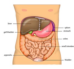
Anatomy Abdomen Tiesworks ku.jpg 674 × 598; 69 KB
-
Anatomy Abdomen Tiesworks zh.jpg 823 × 768; 107 KB
-
Anatomy Abdomen Tiesworks-ar.jpg 642 × 599; 72 KB
-
Anatomy Abdomen Tiesworks.jpg 823 × 768; 210 KB
-
Anatomy and physiology of animals A hair-cs.jpg 585 × 438; 28 KB
-
Anatomy roots Dutch text.jpg 1,281 × 1,561; 224 KB
-
Anatomy The Skin - NCI Visuals Online esp.jpg 1,016 × 1,012; 347 KB
-
Anatomy The Skin - NCI Visuals Online.jpg 1,016 × 1,012; 330 KB
-
Anatomy- bone.jpg 2,359 × 1,800; 794 KB
-
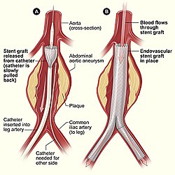
Aneurysm endovascular.jpg 400 × 401; 65 KB
-
Angiospermen - Lebenszyklus.png 2,244 × 2,316; 945 KB
-
Angle facial.png 1,829 × 1,680; 313 KB
-
Animal addtree age7.png 1,360 × 2,191; 97 KB
-
Animal cell diagram.png 360 × 360; 24 KB
-
Antagonismus.png 362 × 296; 8 KB
-
Antena-triatomine.PNG 510 × 1,041; 64 KB
-
Antennae ctb.png 594 × 303; 18 KB
-
Antepassatshome6.jpg 450 × 370; 20 KB
-
Anther morphology attachment basifixed versatile.png 120 × 160; 17 KB
-
Anther morphology attachment basifixed.png 120 × 160; 10 KB
-
Anther morphology attachment dorsifixed versatile.png 120 × 160; 17 KB
-
Anther morphology attachment dorsifixed.png 120 × 160; 16 KB
-
Anther morphology attachment subbasifixed.png 120 × 160; 17 KB
-
Anther morphology dehiscence longitudinal.png 120 × 160; 9 KB
-
Anther morphology dehiscence poricidal.png 120 × 160; 8 KB
-
Anther morphology dehiscence transverse.png 120 × 160; 8 KB
-
Anther morphology dehiscence valvular.png 120 × 160; 10 KB
-
Anther morphology type dithecal.png 120 × 160; 7 KB
-
Anther morphology type monotecal.png 120 × 160; 5 KB
-
Anther-schematic-detail.png 942 × 1,554; 389 KB
-
Anti-HLA agglutinated RBC.png 256 × 294; 11 KB
-
Antibiotic resistance infographic by CDC.jpg 950 × 1,229; 663 KB
-
Antibiotic selection.JPG 1,554 × 4,347; 1.36 MB
-
Antibiotics Mechanisms of action.png 960 × 720; 205 KB
-
Anticossos anticardiolipina.jpg 427 × 286; 61 KB
-
Antisectetory drugs.jpg 2,269 × 2,597; 349 KB
-
Antofiles eix flor.png 400 × 237; 19 KB
-
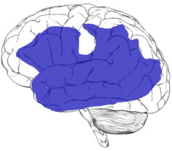
Aphasia.jpg 682 × 599; 83 KB
-
Aphasia.png 682 × 599; 134 KB
-
Apicalconstriction fig1.jpg 417 × 467; 82 KB
-
Apicalconstriction fig2.jpg 393 × 437; 82 KB
-
Apikaldominanz.jpg 322 × 323; 22 KB
-
Apocrine.jpg 749 × 516; 66 KB
-
Apoptose-german.png 1,200 × 1,465; 135 KB
-
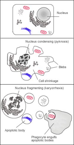
Apoptosis.png 262 × 511; 17 KB
-
Appareil respiratoire vierge.png 800 × 909; 311 KB
-
Appetito.jpg 448 × 600; 120 KB
-
AppoggioPasso.jpg 312 × 587; 156 KB
-
Arbre de parenté des primates.jpg 1,564 × 929; 360 KB
-
Arbre phylogénétique embryophytes.png 1,800 × 1,298; 637 KB
-
Arbre-de-parenté.jpg 1,644 × 828; 509 KB
-
Arm counter-current flow.jpg 1,000 × 528; 121 KB
-
Articulation du genou.jpg 1,506 × 957; 295 KB
-
Artificial Shelter 3a.jpg 783 × 287; 33 KB
-
Ascocarp2.png 666 × 600; 754 KB
-
Astrocyte endothel interaction 01.png 3,156 × 2,578; 718 KB
-
Atheroscleros and artery.png 423 × 599; 27 KB
-
Atherosclerosis diagram.png 450 × 451; 41 KB
-
Atrial fibrillation 01.jpg 800 × 500; 80 KB
-
Atrial Fibrillation in two leads.jpg 434 × 461; 57 KB
-
Atrialfibrillation.jpg 3,038 × 1,521; 2.16 MB
-
.webp.html) Aufbau eines Trägervirus zur Gentherapie (adenoassoziierter Virus, AAV).webp 1,654 × 814; 44 KB
Aufbau eines Trägervirus zur Gentherapie (adenoassoziierter Virus, AAV).webp 1,654 × 814; 44 KB
-
Auge Carybdea marsupialis.jpg 429 × 394; 139 KB
-
Augenstellungen 02.jpg 455 × 245; 40 KB
-
Autopoliploidia.png 809 × 378; 117 KB
-
Autosomal dominant.png 1,058 × 794; 2.41 MB
-
Avermectin PKS.png 22,374 × 6,359; 266 KB
-
Averyversuch.jpg 1,430 × 992; 110 KB
-
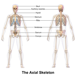
Axial Skeleton.png 2,500 × 2,500; 17.89 MB
-
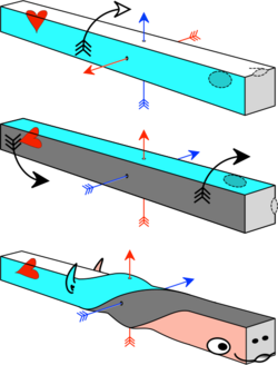
AxialTwistSchema.png 493 × 646; 83 KB
-
B. anthracis Macrophage Invasion Mechanism.jpg 1,856 × 1,012; 167 KB
-
Babesia LifeCycle.png 544 × 435; 41 KB
-
Bacterial colony morphology.png 562 × 564; 39 KB
-
Bacteriophage structure.png 450 × 250; 30 KB
-
Bakterien peritrich Fortbewegung Taumeln.png 587 × 695; 10 KB
-
Bakterien polar begeisselt Schwimmen.png 932 × 400; 6 KB
-
Bakteriophage T2 geschnitten.png 962 × 833; 53 KB
-
Barnaviridae virion.jpg 417 × 248; 63 KB
-
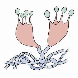
Basidie-2.jpg 1,489 × 1,502; 406 KB
-
Basilic fin vert nain compact Vilmorin-Andrieux 1904.png 3,244 × 3,124; 236 KB
-
Basilic fin vert Vilmorin-Andrieux 1904.png 2,129 × 2,587; 112 KB
-
Basilic grand Vilmorin-Andrieux 1904.png 2,069 × 2,686; 102 KB
-
Basilic à feuille de laitue Vilmorin-Andrieux 1904.png 2,945 × 2,945; 229 KB
-
Batlook3.jpg 655 × 321; 49 KB
-
Bayliss effect.png 503 × 413; 13 KB
-
BBS Organs.jpg 569 × 640; 209 KB
-
Bellis perennis ellywa (cropped).png 4,033 × 5,397; 173 KB